ຄືກັນກັບເລື້ອງການສ່ອງໄຟຟ້າເບິ່ງເຕົ້ານົມ ເພື່ອກວດຫາມະເຮັງ
ເຕົ້ານົມນັ້ນ ກໍມີການໂຕ້ແຍ້ງກັນຫລາຍເຊັ່ນກັນ ກ່ຽວກັບວິທີການ
ກວດເພື່ອວິນິດໄສໂຣກມະເຮັງໃນອັນທະ. ພວກວິຈານກ່າວວ່າ
ທັງສອງວິທີກວດທີ່ນຳໃຊ້ຢູ່ໃນເວລານີ້ ພົບເຫັນເນື້ອງອກນ້ອຍໆ
ທີ່ບໍ່ເປັນອັນຕະ ລາຍ ແຕ່ວ່າສ່ວນຫຼາຍແລ້ວ ບໍ່ພົບເຫັນອັນທີ່
ເປັນອັນຕະລາຍ. ແຕ່ໂຊກດີ ທີ່ມີການສຶກສາຄົ້ນຄວ້າໃໝ່ ທີ່
ສະແດງໃຫ້ເຫັນວ່າ ໃນບໍ່ຊ້ານີ້ ບັນດາທ່ານໝໍອາດມີວິທີກວດ
ທີ່ໜ້າເຊື່ອຖືໄດ້ຫຼາຍຂຶ້ນ ໃນການວິນິດໄສໂຣກມະເຮັງໃນອັນທະ.
ຜູ້ສື່ຂ່າວວີໂອເອ Carol Pearson ມີລາຍງານຕື່ມອີກ ຊຶ່ງ
ໄຊຈະເລີນສຸກ ຈະນຳມາສະເໜີທ່ານໃນອັນດັບຕໍ່ໄປ.
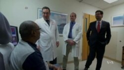
ເບິ່ງຄືວ່າ ໂຣກມະເຮັງໃນອັນທະຫລືຫຳ ຈະເກີດຂຶ້ນກັບທຸກຄົນ ໃນຄອບຄົວ ຂອງທ້າວ Ronald Briscoe.
ທ້າວ Briscoe ເວົ້າວ່າ “ອ້າຍຂອງຂ້ອຍ ພໍ່ຂອງຂ້ອຍ ມີໂຣກນີ້ ແລະ ນ້ອງຫລ້າຂອງຂ້ອຍ ກະມີອີກ.”
ປະຫວັດຂອງຄອບຄົວ ແມ່ນມີສ່ວນກ່ຽວພັນຫຼາຍ ກັບໂອກາດຂອງຜູ້ຊາຍຄົນນຶ່ງ ທີ່ຈະເປັນ
ໂຣກມະເຮັງປະເພດທີ່ອັນຕະລາຍ ໃນອັນທະ.
ການສຶກສາຄົ້ນຄວ້າສະບັບນຶ່ງ ສະແດງໃຫ້ເຫັນວ່າ ຖ້າຫາກຊາຍຄົນນຶ່ງມີຍາດຕິພີ່ນ້ອງຜູ້ຊາຍ ຮວມທັງ ລຸງນ້າບ່າວອາວ ແລະພໍ່ປູ່ພໍ່ຕູ້ ທີ່ເປັນໂຣກມະເຮັງໃນອັນທະນັ້ນ ຄວາມສ່ຽງທີ່ຜູ້ກ່ຽວຈະເປັນໂຣກນີ້ ແມ່ນສາມາດເພີ້ມຂຶ້ນໄດ້ຮອດ 3 ເທົ້າຕົວ.
ບໍ່ຄືກັນກັບໂຣກມະເຮັງໃນເຕົ້ານົມ ຊຶ່ງສາມາດກວດເຫັນໄດ້ດ້ວຍວິທີ ໃຊ້ແສງລັງສີລະດັບຕ່ຳ ສ່ອງເບິ່ງເນື້ອເຫຍື່ອທາງໃນ ທີ່ເອີ້ນວ່າ mammography ນັ້ນ ຕາມປົກກະຕິແລ້ວ ນາຍແພດແມ່ນຈະບໍ່ນຳໃຊ້ວິທີກວດ ທີ່ໃຊ້ເຄື່ອງຖ່າຍພາບທີ່ທັນສະໄໝແບບນີ້ ເພື່ອວິນິດໄສ ໂຣກມະເຮັງໃນຫຳ. ແຕ່ນາຍແພດຈະໃຊ້ວິທີເຮັດ biopsy ກໍຄືໃຊ້ເຂັມເຂົ້າໄປດູດເອົາ ເນື້ອເຫຍື່ອຕົວຢ່າງຂອງອັນທະ ໂດຍໃຊ້ເຄື່ອງ ultrasound ນຳພາເຂັມ. ເຄື່ອງ ultrasound ໃຊ້ຄື້ນສຽງ ຊຶ່ງເປັນເທັກໂນໂລຈີທີ່ປອດໄພ ກວ່າແສງລັງສີ ແຕ່ພວກນາຍແພດກ່າວວ່າ ມັນບໍ່ມີປະສິດທິພາບ ພຽງພໍ.
ດຣ. Peter Pinto ຈາກສະຖາບັນສຸຂະພາບແຫ່ງຊາດສະຫະລັດ ຫລື NIH ເວົ້າວ່າ
“ເຄື່ອງ ultrasound ຮັບປະກັນວ່າ ເຂັມຈະແທງຖືກອັນທະ ແຕ່ພວກເຮົາຈະບໍ່ໃຊ້ ultrasound ເພື່ອນຳພາເຂັມເຂົ້າໄປຫາ ບ່ອນທີ່ອາດມີ ເນື້ອງອກ ນັ້ນ.”
ດຣ. Minhaj Siddiqui ຈາກມະຫາວິທະຍາໄລລັດ Maryland ກ່າວວ່າ ການໃຊ້ເຄື່ອງ ultrasound ນຳພາເຂັມເຂົ້າໄປດູດເອົາເນື້ອເຫຍື່ອຈາກອັນທະນັ້ນ ກໍບໍ່ໄດ້ຜົນສະເໝີໄປ.
ດຣ. Siddiqui ເວົ້າວ່າ “ພວກເຮົາພະຍາຍາມສຸ່ມເລືອກເອົາເນື້ອ ເຫຍື່ອຕົວຢ່າງຂອງອັນທະ ແລະກໍຫວັງວ່າ ຈະແທງຖືກບ່ອນທີ່ມີເນື້ອງອກມະເຮັງ.”
ແຕ່ກົງກັນຂ້າມ ວິທີກວດດ້ວຍເຄື່ອງ MRI ທີ່ໃຊ້ສະໜາມແມ່ເຫຼັກ ແລະຄື້ນວິທະຍຸ ຈະສະແດງໃຫ້ເຫັນພາບລະອຽດຂອງອະໄວຍະວະ ສ່ວນນັ້ນໆ ຂອງຮ່າງກາຍ.
ດຣ. Siddiqui ເວົ້າວ່າ “MRI ສາມາດບອກພວກເຮົາວ່າ ພວກເນື້ອງອກລີ້ຊ່ອນຢູ່ບ່ອນໃດໃນອັນທະ ບ່ອນທີ່ອາດບໍ່ໄດ້ຖືກດູດເອົາອອກມາໂດຍວິທີໃຊ້ເຂັມຕາມປົກກະຕິນັ້ນ.”
ໂຣກມະເຮັງໃນຫຳຂອງທ້າວ Briscoe ນັ້ນ ກໍແມ່ນໄດ້ຖືກກວດເຫັນດ້ວຍເຄື່ອງຖ່າຍພາບ MRI. ຜູ້ກ່ຽວໄດ້ເຂົ້າຮ່ວມການສຶກສາຄົ້ນຄວ້າ ລະດັບໃຫຍ່ ຢູ່ທີ່ສະຖາບັນມະເຮັງແຫ່ງຊາດ ຊຶ່ງເປັນສ່ວນນຶ່ງຂອງ ສະຖາບັນສຸຂະພາບແຫ່ງຊາດສະຫະລັດ. ດຣ. Siddiqui ເປັນຫົວໜ້ານຳພາການສຶກສາຄົ້ນຄວ້ານີ້ ຊຶ່ງພົວພັນກັບພວກຜູ້ຊາຍທີ່ມີຄວາມສ່ຽງສູງ ທີ່ຈະເປັນໂຣກມະເຮັງໃນອັນທະ ຈຳນວນ 1,000 ຄົນ ທີ່ແບ່ງອອກເປັນສອງກຸ່ມ. ກຸ່ມນຶ່ງ ໄດ້ຮັບການກວດຫາເນື້ອເຫຍື່ອທີ່ເປັນມະເຮັງ ດ້ວຍວິທີໃຊ້ທັງເຄື່ອງ ultrasound ແລະ MRI. ສ່ວນອີກກຸ່ມນຶ່ງ ໄດ້ຮັບການກວດ ໂດຍວິທີໃຊ້ເຄື່ອງ ultrasound ນຳພາເຂັມເຂົ້າໄປດູດເນື້ອເຫຍື່ອໃນອັນທະ ຕາມທີ່ເຄີຍເຮັດມາ. ດຣ. Pinto ໄດ້ຮ່ວມຂຽນບົດລາຍງານນີ້ ກັບດຣ. Siddiqui.
ດຣ. Pinto ເວົ້າວ່າ “ການດູດເອົາເນື້ອເຫຍື່ອທີ່ເປັນເປົ້າໝາຍຂອງ ເຮົາ ອອກມາກວດ ໂດຍໃຊ້ເຄື່ອງຖ່າຍ MRI ຊີ້ນຳນັ້ນ ແມ່ນເພີ້ມ ທະວີໂອກາດ ທີ່ຈະກວດເຫັນມະເຮັງໃນຫຳນັ້ນ ຂຶ້ນເຖິງ 30 ເປີເຊັນ ເມື່ອທຽບໃສ່ວິທີການດູດເອົາເນື້ອເຫຍື່ອແບບດັ້ງເດີມ ທີ່ປະຕິບັດກັນ ໃນທຸກມື້ນີ້.”
ເຄື່ອງຖ່າຍ MRI ຊ່ວຍໃຫ້ທ່ານໝໍ ສາມາດແລເຫັນບໍລິເວນເນື້ອເຫຍື່ອ ທີ່ພວກເຂົາເຈົ້າສົງໄສວ່າ ຈະມີເນື້ອງອກແລະຢາກຈະດູດເອົາອອກມາ ກວດເບິ່ງນັ້ນ.
ດຣ. Siddiqui ເວົ້າວ່າ “ປະໂຫຍດໃນການນຳໃຊ້ MRI ກໍແມ່ນວ່າ ມັນເພີ້ມຄວາມສາມາດຂອງພວກເຮົາໃຫ້ສູງທີ່ສຸດ ໃນການທີ່ຈະກວດພົບໂຣກມະເຮັງ ທີ່ມີຄວາມສ່ຽງສູງ ແລະເປັນອັນຕະລາຍ ແລະຍັງອາດຈະຊ່ວຍຫລີກເວັ້ນຄວາມເສຍຫາຍ ຈາກການກວດພົບມະເຮັງ ທີ່ບໍ່ອັນຕະລາຍ ແລະກໍຄົງຈະບໍ່ສ້າງບັນຫາໃດໆ ໃຫ້ຄົນປ່ວຍ.”
ດຣ. Siddiqui ມີແຜນທີ່ຈະທຳການທົດລອງທາງການແພດ ເພີ້ມຕື່ມອີກ ແຕ່ຜົນທີ່ອອກ
ມາໃນຂັ້ນຕົ້ນ ຂອງການຄົ້ນຄວ້ານີ້ ແມ່ນໃຫ້ຄວາມຫວັງສູງ. ລາຍງານການສຶກສາຄົ້ນຄວ້ານີ້
ໄດ້ຖືກພິມລົງໃນວາລະສານ ຂອງສະມາຄົມການແພດອາເມຣິກັນ.
ເບິ່ງວີດີໂອ ກ່ຽວກັບໂຣກມະເຮັງເຕົ້ານົມ ຈະໄດ້ເຫັນວິທີກວດ ທີ່ເອີ້ນວ່າ mammography:
Just like the controversy around mammograms used to find breast cancer, there is a lot of debate about tests used to diagnose prostate cancer.Critics say both procedures find small tumors that should be left alone, and they often fail to find the ones that are dangerous.Fortunately, a new study shows doctors may soon have a more reliable way to diagnose prostate cancer.More from VOA's Carol Pearson.
Prostate cancer seems to run in Ronald Briscoe's family.
"My older brother, my father had it, and also my baby brother has it."
Family history has a lot to do with a man's chances of getting an aggressive type of prostate cancer. One study shows if a man's male relatives had it, his risk could be up to three times higher than normal.
Unlike breast cancer, which can be found with a mammogram using a low dose of radiation, advanced imaging is not normally used to diagnose prostate cancer. Doctors use ultrasound to guide them as they take tissue samples of the prostate.Ultrasound uses sound waves - a safer technology than radiation, but Dr. Peter Pinto from the National Cancer Institute says it falls short of what doctors need.
"Ultrasound insures that the needles hit the prostate, but we do not use the ultrasound to direct the needles into where the tumor may be present."
Dr. Minhaj Siddiqui says biopsies guided by ultrasound are hit or miss.
"We randomly try to get all the prostate sampled in hopes of catching the cancer."
In contrast, an MRI uses a magnetic field and radio waves to give a detailed picture of part of the body.
"MRI can tell us where tumors are hiding in the prostate, that is not normally sampled."
That is how Ronald Briscoe's cancer was found.He participated in a large study at the National Cancer Institute, which is part of the National Institutes of Health.Dr. Siddiqui led the study, which involved 1,000 men at high risk for prostate cancer.One group had a biopsy using both ultrasound and MRI technology.The other group had standard ultrasound-guided biopsies. Dr. Pinto was one of the co-authors.
"The targeted MRI guided biopsy had a 30 percent increase in detection of high-risk prostate cancer as compared to the traditional biopsies performed today."
Dr. Siddiqui plans more clinical trials, but he says results of this preliminary study are promising.
"The benefit of doing that is that we maximize our ability to detect the high risk cancers that matter and also potentially avoid the harm of detecting cancer which is clinically insignificant and unlikely to cause problems for the patient."
This study was published in the journal of the American Medical Association.


