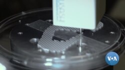
ລອງມາພາກັນວາດພາບເບິ່ງເນາະວ່າ ຊີວິດຂອງຄົນຈະປ່ຽນແປງໄປແບບໃດ ສໍາລັບຫລາຍໆຄົນທີ່ຕ້ອງການປ່ຽນອະໄວຍະວະ ຖ້າບັນດານັກວິທະຍາສາດຫາກສາມາດ ພຽງແຕ່ພິມອະໄວຍະວະອັນໃໝ່ທີ່ໃຊ້ການໄດ້ ອອກມາເທົ່ານັ້ນ. ອັນນັ້ນແມ່ນໂຄງການນຶ່ງໃນຫຼາຍໆໂຄງການທີ່ພວກນັກວິທະຍາສາດຢູ່ສະຖາບັນເວັກຄ໌ ຟໍແຣັສ ສຳລັບການແພດໃນດ້ານການເຮັດໃຫ້ມີຊີວິດຄືນມາໃໝ່ຫລື Wake Forest Institute for Regenerative Medicine ກຳລັງເຮັດຢູ່ແທ້ໆເລີຍ ຊຶ່ງກໍຄືອະໄວຍະວະຂອງມະນຸດທີ່ພິມດ້ວຍເຄື່ອງພິມຊີວະພາບ 3 ມິຕິ ຫລື 3D. ພວກເພິ່ນຍັງພາກັນເຮັດວຽກ ເພື່ອພັດທະນາເສັ້ນເລືອດທຽມ ແລະແບບຈຳລອງຂອງຄົນທີ່ມີຂະໜາດນ້ອຍໆໂດຍໃຊ້ການພິມຊີວະພາບແບບ 3 ມິຕິອີກດ້ວຍ. Lesia Bakalets ສົ່ງລາຍລະອຽດກ່ຽວກັບເລື້ອງນີ້ ມາຈາກເມືອງວິນສຕັນ-ຊາແລມ (Winston-Salem) ລັດຄາໂຣໄລນາເໜືອ ໂດຍຜ່ານນັກຂ່າວຂອງວີໂອເອ Anna Rice, ຊຶ່ງບົວສະຫວັນ ຈະມາສະເໜີທ່ານໃນອັນດັບຕໍ່ໄປ.
ທ້າວ ຈາຊວາ ຄໍພັສ (Joshua Copus), ນັກສຶກສາຂັ້ນປລິນຍາເອກ ເວົ້າວ່າ:
“ພວກເຮົາກຳລັງພິມຫູຂ້າງນຶ່ງ. ສະນັ້ນ, ຄົນປ່ວຍຜູ້ນຶ່ງເສຍຫູຂ້າງຊ້າຍຂອງລາວໄປ. ພວກເຮົາໄດ້ທຳການຖ່າຍຮູບ ຫລື ສະແກນແບບ CT ຫູກ້ຳຂວາຂອງລາວ ແລະເຮັດໃຫ້ມັນຄືກັນເລີຍ ເພື່ອວ່າພວກເຮົາຈະສາມາດພິມຫູໃໝ່ຂອງລາວດ້ວຍແບບຊີວະພາບ 3 ມິຕິ ຫລື 3D ໄດ້.”
ການເອົາມັນຕິດໃສ່ນັ້ນ ຍັງເປັນເລື້ອງເຮັດໄດ້ໃນທາງທິດສະດີຢູ່ ແຕ່ວ່າ ເຄື່ອງພິມ 3 ມິຕິ ຫລື 3D ສາມາດພິມອະໄວຍະວະຂອງຄົນທີ່ໃຊ້ການໄດ້ ເຊັ່ນຫູອັນນີ້.
ພວກນັກວິທະຍາສາດຢູ່ທີ່ນີ້ ໃນສະຖາບັນເວັກຄ໌ ຟໍແຣັສ ສຳລັບການແພດໃນດ້ານການເຮັດໃຫ້ມີຊີວິດກັບຄືນມາ ຢູ່ໃນລັດຄາໂຣໄລນາເໜືອ ກຳລັງດຳເນີນງານ ເພື່ອກ້າວໄປເຖິງວັນເວລາທີ່ພວກເພິ່ນສາມາດພິມອະໄວຍະວະທຸກປະເພດໄວ້ໃຫ້ປ່ຽນແທນໄດ້.
ມັນໃຊ້ເວລາປະມານ 3 ຊົ່ວໂມງເຄິ່ງ ເພື່ອເຮັດຫູອັນນຶ່ງ ແລະມັນຈະຖືກເອົາໄປຕິດໃສ່ໂດຍການໃຊ້ນ້ຳຢາພິເສດອັນນຶ່ງ ທີ່ເຮັດມາຈາກແຊລຂອງຄົນປ່ວຍເອງ.
ເມື່ອພວກເພິ່ນເຮັດໃຫ້ມັນໃຊ້ການໄດ້ແລ້ວ ມັນກໍອາດຈະເປັນການປະຕິວັດວິທະຍາສາດໃນການເອົາອະໄວຍະວະໄປຕິດຕໍ່ໃສ່ກໍເປັນໄດ້.
ທ່ານ ແອນໂທນີ ແອທາລາ (Anthony Atala) ຈາກສະຖາບັນເວັກຄ໌ຟໍແຣັສ ສຳລັບການແພດໃນດ້ານການເຮັດໃຫ້ມີຊີວິດກັບຄືນມາ ກ່າວວ່າ:
“ພວກເຮົາໂດຍພື້ນຖານແລ້ວ ຈັດປະເພດຄວາມສັບສົນຂອງອະໄວຍະວະນັບ ແຕ່ອັນທີ່ພຽງໄປເຖິງອັນທີ່ແໜ້ນ. ສຳລັບໂຄງສ້າງອັນທີ່ພຽງ, ເຊັ່ນຜິວໜັງ ແມ່ນມີໂຄງສ້າງທີ່ສັບສົນໜ້ອຍທີ່ສຸດ, ໂຄງສ້າງເປັນທໍ່ ເຊັ່ນເສັ້ນເລືອດ ແມ່ນມີຄວາມສັບສົນໃນລະດັບສອງ, ອັນທີ່ໂຄ່ງທີ່ບໍ່ແມ່ນອະໄວຍະວະທີ່ເປັນຮູບທໍ່ເຊັ່ນ ພົກປັດສະວະ ຫລື ກະເພາະອາຫານແມ່ນ ເປັນອະໄວຍະວະທີ່ມີຄວາມສັບສົນອັນດັບ 3 ແລະອະໄວຍະວະທີ່ແຂງ ແມ່ນອະໄວຍະວະທີ່ມີຄວາມສັບສົນຫຼາຍ ກວ່ານັ້ນຫລາຍ. ແນ່ນອນ ການຂາດເຂີນຜູ້ບໍລິຈາກອະໄວຍະວະ ເປັນບັນຫາທີ່ຮ້າຍແຮງຫລາຍ. ທ່ານຮູ້ບໍ່ ມີຄົນປ່ວຍຫລາຍໆຄົນ ຢູ່ໃນບັນຊີຂອງຄົນລໍຖ້າຢູ່ດຽວນີ້ ແລະການແພດທີ່ເຮັດໃຫ້ມີຊີວິດໃໝ່ກັບຄືນມາອີກ ແມ່ນເປັນຍຸດທະສາດທີ່ຫວັງວ່າ ຈະຊ່ວຍພວກເຮົາຫລຸດຜ່ອນຈຳນວນຄົນປ່ວຍທີ່ຢູ່ໃນບັນຊີຂອງຄົນລໍຖ້າປ່ຽນອະໄວຍະວະນັ້ນ ລົງໄດ້ອີ່ຫລີ.”
ຂັ້ນຕອນແມ່ນເລີ້ມຕົ້ນຂຶ້ນເມື່ອພວກນັກວິທະຍາສາດ ເອົາເນື້ອເຫຍື່ອນ້ອຍໆຕ່ອນນຶ່ງ ຈາກຄົນປ່ວຍໄປ. ຂັ້ນຕອນຕໍ່ໄປກໍຄື ເຮັດໃຫ້ເນື້ອເຫຍື່ອນັ້ນ ເປັນຮູບຮ່າງ ຫລື ໂຄງປະກອບທີ່ພວກເຮົາຢາກໄດ້. ນີ້ກໍຄືບ່ອນທີ່ເຄື່ອງພິມແບບ 3 ມິຕິ ເຂົ້າມາມີບົດບາດ ແລະ ຢູ່ຄຽງຂ້າງພວກມັນນັ້ນກໍຄືຂັ້ນຕອນທີ່ເອີ້ນວ່າ ວິທີຜະລິດເສັ້ນໃຍດ້ວຍການປັ່ນທີ່ໃຊ້ພະລັງງານໄຟຟ້າ ເພື່ອເຮັດເສັ້ນເລືອດທຽມ.
ທ່ານເດວິດ ແມັກຄາວລ໌, ນັກຄົ້ນຄວ້າທີ່ໄດ້ຮັບທຶນໄປຄົ້ນຄວ້າຢູ່ສະຖາບັນແຫ່ງ ນັ້ນ ໃຫ້ຄຳເຫັນວ່າ:
“ເຄື່ອງເຫລົ່ານັ້ນຄື ເຄື່ອງປັ່ນໃຊ້ພະລັງງານໄຟຟ້າ. ແລະ ວິທີທີ່ມັນທຳງານກໍຄື ພວກເຮົາມີນ້ຳຢາ ຈາກວັດຖຸຊີວະພາບ, ສານໂພລີເມີຊະນິດໃດນຶ່ງຢູ່ໃນຫລອດເຂັມສີດຢາທີ່ຍູ້ອອກໄປຢ່າງຊ້າໆ ດ້ວຍການໃຊ້ເຄື່ອງດູດໃນຫລອດເຂັມສີດຢາ. ພວກເຮົາມີເສັ້ນລວດຢູ່ໜີ້ທີ່ສອດຜ່ານເຄື່ອງມືອັນນີ້ ຢູ່ບ່ອນໜີ້ ແລະມັນກໍໝູນວຽນໄປມາ ແລະເໜັງໄປ ເໜັງມາ. ແລະຄວາມຄິດກໍຄື ນ້ຳຢານັ້ນແມ່ນເອົາຕິດໃສ່ເສັ້ນລວດ ແລະມັນກໍສາມາດທີ່ຈະເຮັດໃຫ້ເກີດເປັນທໍ່ຂຶ້ນມາໄດ້.”
ຕໍ່ໄປ, ພວກນັກວິທະຍາສາດກໍສາມາດທົດລອງເສັ້ນເລືອດທຽມໄດ້ໂດຍການກ່າຍເອົາແບບວິທີເຕັ້ນຂອງຊີບພະຈອນ.
ທ່ານເດວິດ ແມັກຄາວລ໌ ອະທິບາຍຕໍ່ໄປອີກວ່າ:
“ພວກເຮົາເອົາມັນໄປໄວ້ໃນເຄື່ອງປະຕິກອນ ຫລືເຄື່ອງກອງຊີວະພາບ. ສິ່ງທີ່ ພວກເຮົາໄດ້ເຮັດກໍຄື ພວກເຮົາໄດ້ເອົາເສັ້ນເລືອດຢູ່ນີ້ໜີ້, ພວກເຮົາໄດ້ເອົາມັນໄປຕິດໃສ່ກັບເຄື່ອງປະຕິກອນຊີວະພາບທີ່ຕິດຢູ່ກັບເຄື່ອງປ້ຳອັນນຶ່ງ. ແລະໂດຍພື້ນຖານແລ້ວ ມັນກໍເຕັ້ນເປັນຈັງຫວະເຮັດໃຫ້ເລືອດໄຫລກັບໄປກັບມາ ຜ່ານເສັ້ນເລືອດເພື່ອເຮັດໃຫ້ເປັນຄືກັນກັບການເຕັ້ນຂອງຫົວໃຈ. ແລະເພາະສະນັ້ນ ທ່ານກໍສາມາດເຫັນ ເສັ້ນເລືອດເປີດຂຶ້ນ ແລະປິດລົງຢູ່.”
ຍ້ອນວ່າມີນັກວິທະຍາສາດດ້ານການພິມຊີວະພາບແບບ 3 ມິຕິ ສາມາດສ້າງອະໄວຍະວະມະນຸດ ທີ່ສັບສົນຫລາຍກວ່ານີ້ໄດ້ອີກດ້ວຍ ພວກອະໄວຍະວະນີ້ ຈຶ່ງຖືກໃຊ້ຢູ່ໃນເທັກໂນໂລຈີ ທີ່ເອີ້ນກັນວ່າ ອະໄວຍະວະຢູ່ເທິງປ່ຽງນ້ອຍໆ ຫລື OCP.
ແຜງທີ່ກ້ວາງ 5 ຊັງຕີແມັດນີ້ ເປັນແບບຈຳຮອງຂອງຮ່າງກາຍຄົນ. ແຕ່ລະຫ້ອງ ແມ່ນບັນຈຸເນື້ອເຫຍື່ອຈາກອະໄວຍະວະຫລາຍອັນ. ແບບຈຳຮອງເຊັ່ນນັ້ນ ແມ່ນເກືອບວ່າໃຊ້ເພື່ອທົດລອງຢາໃໝ່ສຳລັບປົວຜົນຂ້າງຄຽງທີ່ບໍ່ມີໃຜຮູ້ ແລະແມ່ນແຕ່ເພື່ອເບິ່ງວ່າ ມັນຈະມີຜົນກະທົບຕໍ່ຄົນປ່ວຍແຕ່ລະຄົນຄືແນວໃດອີກດ້ວຍ.
ທ່ານ ຈູລຽວ ອາເລແມນ, ນັກຄົ້ນຄວ້າທີ່ໄດ້ຮັບທຶນໄປຄົ້ນຄວ້າຢູ່ສະຖາບັນແຫ່ງ ນັ້ນ ໃຫ້ຄຳເຫັນວ່າ:
“ໃນອະນາຄົດອັນໃກ້ໆນີ້ ສິ່ງທີ່ຈະເກີດຂຶ້ນ ກໍຄື ທ່ານຈະມາຫາໂຮງໝໍ ແລ້ວ ພວກເຂົາເຈົ້າ ກໍຈະເອົາຜິວໜັງຂອງທ່ານໜ້ອຍນຶ່ງເພື່ອໄວ້ເປັນຕົວຢ່າງ ແລະໃນຂະນະທີ່ພວກທ່ານໝໍ ກຳລັງວິໄຈເພື່ອທຳນາຍເບິ່ງອາການໂຣກຂອງທ່ານ ແລະທຸກສິ່ງທຸກຢ່າງຢູ່ນັ້ນ ກຸ່ມຫ້ອງທົດລອງກໍຈະສ້າງແບບສະບັບຂອງທ່ານ 20 ຫາ 40 ແບບ ຂຶ້ນມາຢູ່ເທິງຊິບ ຫຼືປ່ຽງນ້ອຍໆ ຫລາຍອັນ."
ພວກນັກວິທະຍາສາດ ບໍ່ສາມາດບອກໄດ້ວ່າ ການທົດລອງຢູ່ໃນຄລີນິກ ຈະໃຊ້ເວລາດົນປານໃດ ແຕ່ພວກເພິ່ນກໍຫວັງວ່າ ໃນປະມານ 30 ຫາ 40 ປີຂ້າງໜ້ານີ້ ການປະດິດຄິດສ້າງຂອງພວກເພິ່ນຈະເປັນສ່ວນນຶ່ງທີ່ປົກກະຕິຢູ່ໃນຊີວິດປະຈຳວັນຂອງຄົນໄດ້.
ອ່ານລາຍງານນີ້ເພີ້ມເປັນພາສາອັງກິດຢູ່ລຸ່ມນີ້
Imagine how life would change for so many people who need a replacement organ if scientists could just print a new one that works. That's exactly one of the projects scientists at Wake Forest Institute for Regenerative Medicine are working on - 3D bio printed human organs. They're also working to develop artificial blood vessels and miniature models of a human body using 3d bio printing. Lesia Bakalets has the story from Winston-Salem, North Carolina in this report narrated by Anna Rice.
Joshua Copus, Graduate Student:
We're printing an ear. So, a patient had lost his left ear. They took a CT scan of his right ear and mirrored it so that we can 3D bio print him a new ear."
Attaching them is still theoretical, but these 3D printers can print workable human organs, like this ear.
Scientists here at the Wake Forest Institute for Regenerative Medicine in North Carolina, are working towards the day when they can print all kinds of replacement organs.
It takes about three and a half hours to create an ear, and it will be attached using a special solution made from the patients cells.
When they get it to work, it could revolutionize the science of organ transplants.
Anthony Atala, Director, Wake Forest Institute for Regenerative Medicine:
We basically categorize the complexity of the organs from flat to solid. With flat structures, such as skin being the least complex, tubular structures like blood vessels being the second level of complexity; hollow, not tubular organs, like the bladder or the stomach being the third level and solid organs being by far the most complex. //Certainly the donor shortage is very serious. You know there's so many patients on the waitlist right now and regenerative medicine is really a strategy that hopefully will help us to decrease the number of patients who are on that transplant list."
The process begins when scientists take a small piece of tissue from the patient. The next step is to create the desired shape or frame for the tissue. This is where 3D printers come into play, and alongside them the procedure known as electrospinning a fiber production method which uses electric force to create artificial blood vessels.
David McCoul, Institute Research Fellow:
These are electrospinners. And the way that it works is we have a solution of bio material a certain type of a polymer in a syringe that's being very slowly pushed out with a syringe pump. // We have a rod here that goes through this apparatus over here and it rotates and moves back and forth. And the idea is that the solution is deposited onto the rod and it can form a tube."
Next, scientists test the artificial blood vessel by imitating a pulse.
David McCoul, Institute Research Fellow:
“We put it inside a bioreactor. What we've done is we've taken the blood vessel right here, we've hooked it up to the bioreactor, which is attached to a pump. And it's basically pulsing flow back and forth through the blood vessel to emulate the beating of the heart. And so you can see the blood vessel is opening and closing."
Thanks to 3D bio printing scientists can create more complex human organs as well they are used in the so-called organ-on-a-chip, or OCP, technology.
This 5-centimeter panel is a tiny model of a human body. Each chamber contains tissues from various organs. Such models are mostly used for testing new drugs for unknown side effects and even how they'll effect each patient individually.
Julio Aleman, Institute Research Fellow:
In the near future what is going to happen is you're going to come to the hospital, they are going to take a small sample from your skin and as the doctor is making your diagnosis on your prognosis and everything, a lab group is going to be building 20 to 40 versions of you in chips."
Scientists can't say how long clinical trials are going to take exactly, but they hope that in some 30-40 years their inventions will be a regular part of people's everyday life.


